Elbow dysplasia is a developmental disease affecting the elbows of dogs and is an umbrella term encompassing fragmented medial coronoid process (also known as coronoid disease and often abbreviated to FCP), ununited anconeal process (UAP) and osteochondrosis of the medial humeral condyle (OCD).
Fragmented medial coronoid process is by far the most common manifestation of the condition.
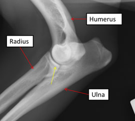
A visually normal elbow joint. The yellow arrow indicates the medial coronoid process of the ulna which overlaps with the radius in this view
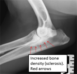
This image shows sclerosis which is indicative of fragmented medial coronoid process
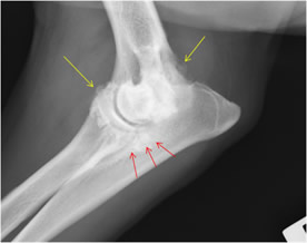
Chronic osteoarthritis secondary to elbow dysplasia. Yellow arrows show new bone.
Red arrows show increased bone density (sclerosis)
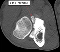
Elbow CT (computed tomography) scan
How does elbow dysplasia happen?
Elbow dysplasia is an inherited (genetic) problem however the exact sequence of events that produces these changes has not been fully worked out.
It is thought that malalignment of the three bones (humerus, radius and ulna) which make up the elbow joint is the main problem but this is not appreciable in every case at the time of diagnosis.
What are the symptoms?
The first thing that is usually seen is stiffness after rest or exercise with some dogs lame at all times.
How do we diagnose it?
A combination of examination, x-ray, computed tomography (CT), arthroscopy.
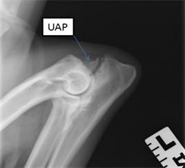
Ununited anconeal process
What can be done about it?
Conservative Management
A proportion of dogs will recover with exercise restriction, pain relieving medication combined with weight loss if appropriate.
We generally advise at least four weeks of this prior to any surgical intervention.
Arthroscopy
Arthroscopy is a method of ‘keyhole’ surgery and is used to evaluate the joint, remove any loose fragments of bone and cartilage and document cartilage damage and loss.
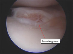
Fragmented medial coronoid process with loose fragment
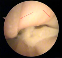
Advanced cartilage loss secondary to elbow dysplasia. Arrows indicate grooves eroded into underlying bone
Osteotomy
Osteotomy means cutting a bone. Several methods have been described for the treatment of elbow dysplasia. The aim is either to allow the joint to become better aligned or to change the weight bearing axis of the limb which means that the main forces are distributed through an area of the joint other than the damaged area.
Objective evidence is lacking in their efficacy and this is rarely our first line of treatment. It is reserved for cases with a significant joint malalignment.
Joint replacement
Elbow replacement can be performed in dogs however it is in the fairly early stages of use in clinical cases. There are two main types which are partial and full joint replacement.
At the moment the developers of both procedures report a significant complication rate and this treatment is reserved for only the most severely affected cases.
What is the outcome?
Frustratingly this is a condition that we cannot reverse once it is present however it is possible to improve matters in the majority of cases.
A general prognosis for fragmented medial coronoid process taken from the largest available study for dogs treated arthroscopically found that in 60% lameness resolves, in 30% lameness improves but does not resolve and 10 % have no improvement. Osteoarthritis progresses in about 70% of cases.
Prevention
When buying a puppy from a breed commonly affected by the condition such as Labradors, Rottweilers, Mastiffs, etc do your homework and always make sure that both parents have been elbow scored with a 0/0 score.
Although this does not guarantee that the condition will not occur, it makes it much less likely.
© Andrew Miller & Associates 2016
Thank you to everyone at Broadley’s, especially Luke. My wee dog Rosie has had TPLO surgeries done on both her back legs, since the beginning of the year.
This has made such a difference to her mobility and she has started to enjoy playing with her toys again and going for her wee walk. Rosie has received excellent care from Broadley’s, the nurses and reception have been so helpful when we have been at the hospital and with Rosie’s aftercare.
Luke has been fantastic with Rosie and with his expertise, advice and the operations he did for Rosie, she is now pain free and is a happy wee dog again enjoying life.
I am truly grateful to Luke and the staff at Broadley’s for what they have done for Rosie.
Marie Terese Xx
Our dog Lady was seen by Andrew.
Very grateful for all care given to our furbaby and counting down the weeks until she can be off the lead and running around crazy again.
We hope not to see you again but know that if we do our girl will be great hands. The photo of Lady shows her with a big smile at being able to go swimming again because of Andrew’s fantastic work.
Donna
Our beagle Alfie was recommended to go here from our own vets in Glasgow as Alfie needed surgery on his leg.
So we met with one of the vet surgeons who took Alfie in for xrays n spoke to us about the options. Andrew n his team completed both parts of the TPLO surgery on Alfie around 2 and a bit years ago.
I’d like to say thank u to everyone in Broadleys for helpin us arrange the stuff with the insurance company and lookin after my dog…. Alfie is now happy and ud never know he had the surgery got a lot mire energy in him now!
Mandy
Orthopaedic Referral Service
Our orthopaedic service, with state-of-the-art facilities, including MRI and CT scanning, and expert nursing care, provides the highest level of veterinary orthopaedic care available.
We use the highest quality equipment and perform minimally invasive techniques where appropriate from Chihuahuas to Great Danes.
We have special miniature fracture systems for cats and small dogs.